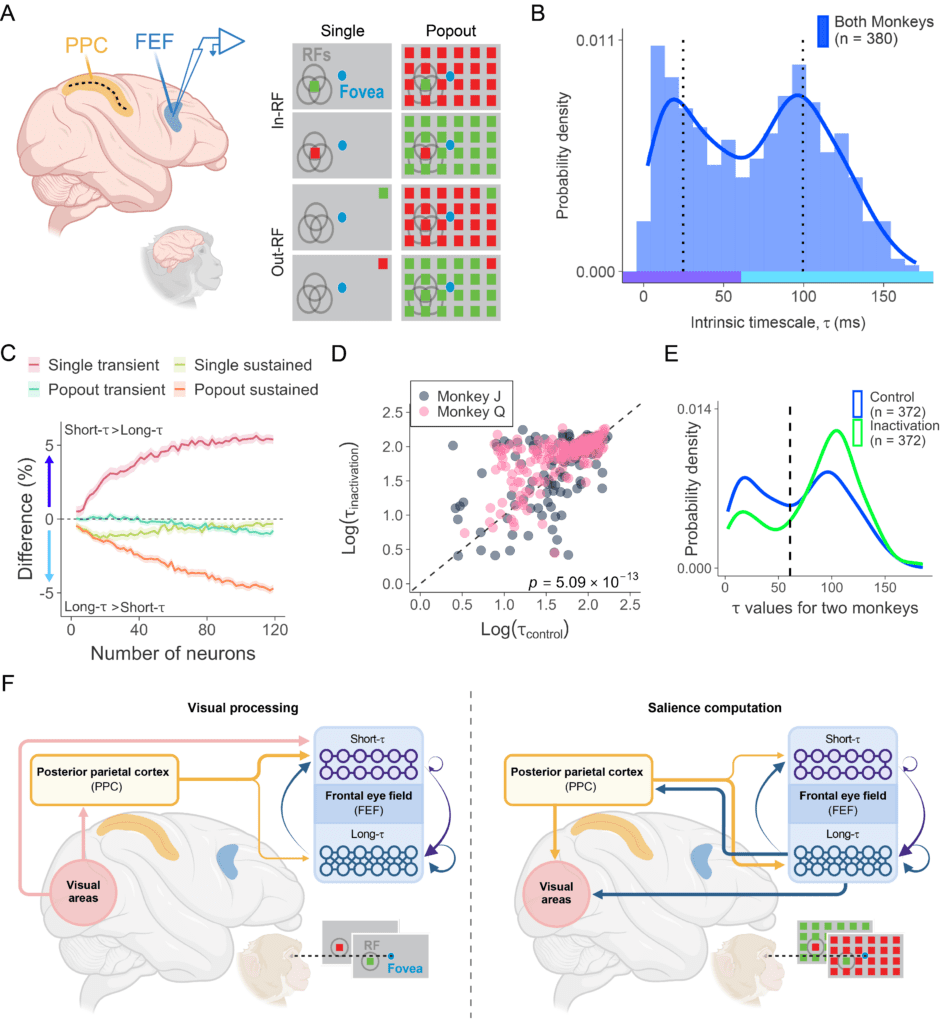
The frontal eye field (FEF) and posterior parietal cortex (PPC) are key components of the brain’s attention network, but how parietal input shapes prefrontal neural dynamics has remained unclear. In this study, we recorded from FEF neurons in macaques and found two distinct populations with different intrinsic timescales: short-timescale neurons, which were more strongly involved in rapid visual processing, and long-timescale neurons, which showed stronger sustained representation of visual salience. By inactivating PPC, we found causal evidence that parietal input regulates these prefrontal dynamics: neural timescales in FEF became slower overall, with larger effects in short-timescale neurons, while salience representation was selectively disrupted, especially in long-timescale neurons. These findings show that long-range interactions between parietal and prefrontal cortex shape the temporal dynamics that support visual attention.
Soyuhos, O., Zirnsak, M., Chaudhuri, R., & Chen, X. (2026). Selective control of prefrontal neural timescales by parietal cortex. Nature Communications, 17, 3687. https://doi.org/10.1101/2024.09.30.615928.
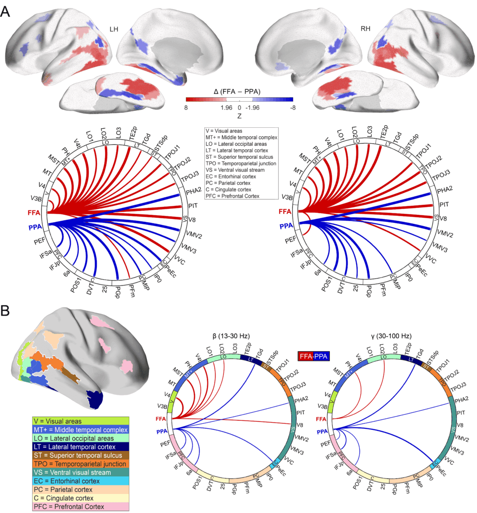
Face and scene perception rely on specialized cortical regions, but how these regions are embedded within larger intrinsic brain networks remains unclear. Using resting-state fMRI and MEG, we found that the fusiform face area (FFA) and parahippocampal place area (PPA) anchor distinct functional connectomes with different spatial and frequency-specific profiles. The FFA network was more strongly linked to lateral occipitotemporal, inferior temporal, and temporoparietal regions, whereas the PPA network was more strongly linked to ventromedial visual, posterior cingulate, and entorhinal-perirhinal regions. This segregation was also reflected in beta- and gamma-band MEG connectivity. Importantly, connectome-based predictive modeling revealed a double dissociation: intrinsic connectivity within the FFA network predicted performance on a face-matching task, while intrinsic connectivity within the PPA network predicted performance on scene tasks, but not vice versa. These findings show that face and scene perception are supported by distinct intrinsic large-scale networks whose architecture is linked to individual differences in behavior.
Soyuhos, O., Scarpa, A., & Baldauf, D. (2026). Distinct resting-state connectomes for face and scene perception predict individual task performance. Human Brain Mapping, 47(5), e70498. https://doi.org/10.1002/hbm.70498.
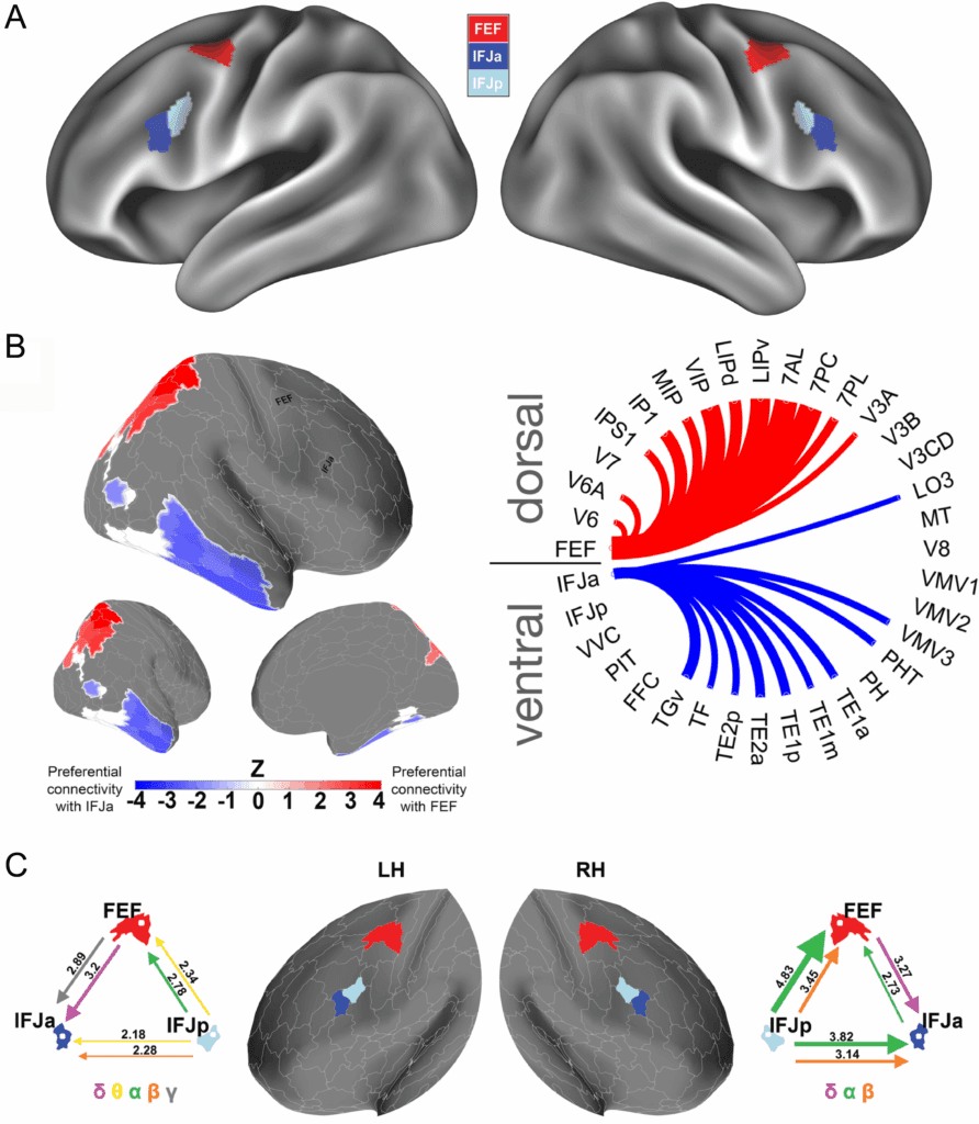
The frontal eye field (FEF) and inferior frontal junction (IFJ) are both key regions in attention and working memory, but their distinct large-scale connectivity patterns had not been directly compared. Using resting-state MEG and seed-based functional connectivity analyses, we found a clear dissociation between these two prefrontal regions: FEF was more strongly coupled to the dorsal visual stream, especially superior parietal and intraparietal regions, whereas anterior IFJ (IFJa) was more strongly coupled to ventral visual and temporal regions associated with feature- and object-based processing. These interactions were also frequency-specific, with FEF showing stronger coupling to dorsal regions particularly in beta-band activity, while IFJa showed stronger coupling to ventral regions especially in delta and gamma bands. Together, these findings show that the prefrontal cortex carries distinct dorsal and ventral connectivity fingerprints, consistent with spatial versus nonspatial processing in attention and working memory.
Soyuhos, O., & Baldauf, D. (2023). Functional connectivity fingerprints of the frontal eye field and inferior frontal junction suggest spatial versus nonspatial processing in the prefrontal cortex. European journal of neuroscience, 57(7), 1114–1140. https://doi.org/10.1111/ejn.15936.